・Tateyama H, Sugiura H, Yamatani C, Yano M.
Expression of podoplanin in thymoma: its correlation with tumor invasion, nodal metastasis, and poor clinical outcome.
Hum Pathol. 2011 Jan 13. [Epub ahead of print]
*我々の論文2報(Kato, Tumor Biol. 2005;Kunita, AJP. 2007)が引用されている。
Abstract
Recent studies have shown that podoplanin overexpression is associated with lymph node metastasis and poor clinical outcome in several malignant tumors. To investigate the role of podoplanin in thymoma, we examined 111 thymomas by immunohistochemistry using monoclonal antibody D2-40, which recognizes podoplanin. The tumors consisted of 8 type A, 40 type AB, 15 type B1, 23 type B2, 15 type B3, and 10 combined thymomas according to the World Health Organization histological classification system and of 41 stage I, 28 stage II, 16 stage III, 20 stage IVa, and 6 stage IVb thymomas according to the Masaoka staging system. We have found podoplanin expression in 0 (0%) type A, 4 (10%) type AB, 4 (27%) type B1, 16 (70%) type B2, 10 (67%) type B3, and 7 (70%) combined thymomas and in 5 (12%) cases of stage I, 7 (25%) of stage II, 11 (69%) of stage III, 12 (60%) of stage IVa, and all (100%) of stage IVb thymomas. Podoplanin was significantly expressed in B2/B3/combined thymomas and advanced stage thymomas (P < .0001). On survival analysis, podoplanin expression was significantly associated with an increased risk of death for the whole group of thymomas (P = .0002), although it was not identified as an independent prognostic factor in multivariate analysis. The significant survival curve differences of podoplanin expression were also seen for stage III/IVa/IVb thymomas (P = .0409) and B2/B3/combined thymomas (P = .0478). In conclusion, D2-40 immunostaining seems to be valuable for predicting the aggressive and metastatic potential of thymomas and the prognosis of the patients.
・Xu Y, Ogose A, Kawashima H, Hotta T, Ariizumi T, Li G, Umezu H, Endo N.
High-level expression of podoplanin in benign and malignant soft tissue tumors: Immunohistochemical and quantitative real-time RT-PCR analysis.
Oncol Rep. 2011 Jan 13. doi: 10.3892/or.2011.1141. [Epub ahead of print]
*我々の論文9報(Kato, Tumor Biol. 2005; Suzuki-Inoue, JBC 2007; Kato, Cancer Sci. 2008; Kaneko, FEBS lett. 2007; Kato, JBC 2003; Kato, BBRC 2006; Mishima, ANP 2006a, Mishima, ANP 2006b; Suzuki, FEBS lett 2008)が引用されている。
Abstract
Podoplanin is a 38 kDa mucin-type transmembrane glycoprotein that was first identified in rat glomerular epithelial cells (podocytes). It is expressed in normal lymphatic endothelium, but is absent from vascular endothelial cells. D2-40 is a commercially available mouse monoclonal antibody which binds to an epitope on human podoplanin. D2-40 immunoreactivity is therefore highly sensitive and specific for lymphatic endothelium. Recent investigations have shown widespread applications of immunohistochemical staining with D2-40 in evaluating podoplanin expression as an immunohistochemical marker for diagnosis and prognosis in various tumors. To determine whether the podoplanin (D2-40) antibody may be useful for the diagnosis of soft tissue tumors, 125 cases, including 4 kinds of benign tumors, 15 kinds of malignant tumors and 3 kinds of tumor-like lesions were immunostained using the D2-40 antibody. Total RNA was extracted from frozen tumor tissue obtained from 41 corresponding soft tissue tumor patients and 12 kinds of soft tissue tumor cell lines. Quantitative real-time PCR reactions were performed. Immunohistochemical and quantitative real-time RT-PCR analyses demonstrated the expression of the podoplanin protein and mRNA in the majority of benign and malignant soft tissue tumors and tumor-like lesions examined, with the exception of alveolar soft part sarcoma, embryonal and alveolar rhabdomyosarcoma, extraskeletal Ewing's sarcoma/peripheral primitive neuro-ectodermal tumor and lipoma, which were completely negative for podoplanin. Since it is widely and highly expressed in nearly all kinds of soft tissue tumors, especially in spindle cell sarcoma, myxoid type soft tissue tumors and soft tissue tumors of the nervous system, podoplanin is considered to have little value in the differential diagnosis of soft tissue tumors.
・Suzuki H, Onimaru M, Koga T, Takeshita M, Yano T, Maehara Y, Nakamura S, Sueishi K.
High podoplanin expression in cancer cells predicts lower incidence of nodal metastasis in patients with lung squamous cell carcinoma.
Pathol Res Pract. 2010 Dec 29. [Epub ahead of print]
*我々の論文3報(Suzuki-Inoue, JBC 2007, Nakazawa Blood 2008; Kunita, AJP 2007)が引用されている。
Podoplanin is also able to induce platelet activation/aggregation mediated by its platelet aggregation-stimulating (PLAG) domain, causing greater ability to achieve hematogenous metastasis of circulating tumor cells in animal models [10,15,23].
抗ポドプラニン抗体として、AngioBioから販売されているマウス抗ポドプラニン抗体(クローン:D2-40)を使用。(AngioBioからは正式に公表されていないが、AngioBioから販売されているマウス抗ポドプラニン抗体(Cat No.:11-003)はD2-40である。)
Abstract
Podoplanin is expressed in a variety of malignant cells, and is generally regarded as a factor promoting tumor progression in conventional studies. Conversely, a recent clinicopathological study has revealed that low podoplanin in cancer cells was correlated with poor prognosis of patients with stage IB lung squamous cell carcinoma (LSCC). We here evaluated the clinicopathological relationship between cancer-cell podoplanin expression and clinicopathological parameters in 40 cases of LSCC (stage I-III). Immunohistochemical podoplanin expression significantly correlated with N classification and pathological stage, but not with other clinicopathological parameters. Notably, all 16 cases with high podoplanin expression unexceptionally exhibited pathological N0 status. Cases without nodal metastasis showed a significantly higher podoplanin-positive score. Furthermore, patients with high podoplanin expression exhibited a significantly longer survival time and disease-free time. These findings suggest that immunohistochemical analysis for podoplanin may serve as a marker of risk of nodal metastasis and prognosis in patients with LSCC.
Int J Clin Exp Pathol. 2011 Jan 30;4(2):175-82.
D2-40: an additional marker for myoepithelial cells of breast and the precaution in interpreting tumor lymphovascular invasion.
Ren S, Abuel-Haija M, Khurana JS, Zhang X.
Department of Pathology and Laboratory Medicine, Temple University Hospital, Temple University School of Medicine Philadelphia, PA 19140, USA.
Abstract
D2-40 is a recently available mouse monoclonal antibody specific for human podoplanin and has been used in identifying lymphovascular invasion (LVI) of tumors. Although its expression has been evaluated in other tissues, its use as a marker for myoepithelial cells (MEC) of breast has not been studied. To explore its expression in the MEC of breast, paraffin-embedded tissue blocks from 48 patients with breast diseases were selected to include usual ductal hyperplasia (UDH, 41 cases), atypical ductal hyperplasia (ADH, 4 cases) and ductal carcinoma in situ (DCIS, 17 cases). Normal breast parenchyma and invasive carcinoma coexisting in the tissue sections were also included in the study. Immunohistochemistry for D2-40, calponin and p63 was performed and the staining patterns were reviewed and compared. D2-40 immunohistochemical staining is positive in the cytoplasm of MEC in UDH, ADH, and the majority of DCIS. The staining pattern of D2-40 is comparable with that of calponin, however D2-40 staining of MEC is weaker than that of calponin and with less background. In addition, myoepithelial cells and myofibroblasts at the edge of retraction spaces of DCIS are also stained by D2-40 that could be misinterpreted as tumor LVI. In conclusion, D2-40 immunohistochemistry reliably identifies the MEC of breast in a variety of lesions in a pattern similar to that of calponin and p63, and can be used as an additional MEC marker. Caution should be exercised when interpreting the staining of cells surrounding DCIS and carcinoma with retraction artifact.
PMID: 21326813 [PubMed - in process]
・Fernandez-Munoz B, Yurrita MM, Martin-Villar E, Carrasco-Ramirez P, Megias D, Renart J, Quintanilla M.
THE TRANSMEMBRANE DOMAIN OF PODOPLANIN IS REQUIRED FOR ITS ASSOCIATION WITH LIPID RAFTS AND THE INDUCTION OF EPITHELIAL-MESENCHYMAL TRANSITION.
Int J Biochem Cell Biol. 2011 Mar 2. [Epub ahead of print]
*我々の論文1報(Kunita, AJP. 2007)が引用されている。
Abstract
Podoplanin is a transmembrane glycoprotein that is upregulated in cancer and was reported to induce an epithelial-mesenchymal transition (EMT) in MDCK cells. The promotion of EMT was dependent on podoplanin binding to ERM (ezrin, radixin, moesin) proteins through its cytoplasmic (CT) domain, which led to RhoA-associated kinase (ROCK)-dependent ERM phosphorylation. Using detergent-resistant membrane (DRM) assays, as well as transmembrane (TM) interactions and ganglioside GM1 binding, we present evidence supporting the localization of podoplanin in raft platforms important for cell signalling. Podoplanin mutant constructs harbouring a heterologous TM region or lacking the CT tail were unable to associate with DRMs, stimulate ERM phosphorylation and promote EMT or cell migration. Similar effects were observed upon disruption of a GXXXG motif within the TM domain, which is involved in podoplanin self-assembly. In contrast, deletion of the extracellular (EC) domain did not affect podoplanin DRM association. Together, these data suggest that both the CT and TM domains are required for podoplanin localization in raft platforms, and that this association appears to be necessary for podoplanin-mediated EMT and cell migration.
ポドプラニンの免疫測定法
概要
肺癌及び悪性中皮腫の検査において従来の免疫染色で必須であった組織採取を必要とせず、患者の負担軽減を可能とし、肺癌及び悪性中皮腫の指標となるデータを収集する方法を提供することにある。 体液中のポドプラニンを測定することを特徴とするポドプラニンの免疫測定法。該測定法においてポドプラニンに対するモノクローナル抗体を用いることが好ましく、特にモノクローナル抗体が、細胞膜表面から外側に突出しているポドプラニンの細胞外領域を認識する抗体及びポドプラニンの細胞外領域以外を認識する抗体を含むことが好ましい。また、前記体液は血液又は胸水であることが好ましく、前記ポドプラニンは肺癌又は中皮腫に関連するポドプラニンであることが好ましい。
目的
本発明の課題は、肺癌及び悪性中皮腫の検査において従来の免疫染色で必須であった組織採取を必要とせず、患者の負担軽減を可能とし、肺癌及び悪性中皮腫の指標となるデータを収集する方法を提供することにある
効果
本発明の測定方法により、肺癌及び悪性中皮腫の検査で、従来の免疫染色で必要であった組織採取が必要でなく、患者の負担を軽減できる。
・Hoshino A, Ishii G, Ito T, Aoyagi K, Ohtaki Y, Nagai K, Sasaki H, Ochiai A.
Podoplanin-positive fibroblasts enhance lung adenocarcinoma tumor formation.
Cancer Res. 2011 May 24. [Epub ahead of print]
*我々の論文1報(Kunita, AJP. 2007)が引用されている。
Abstract
During the metastatic process, cancer cells beleaguer and interact with vascular adventitial fibroblasts (VAFs), which are the main components of the outermost connective tissue layer of blood vessels. This activity suggests the presence of a specific tumor microenvironment in the perivascular area. The subcutaneous co-injection of human lung adenocarcinoma cell lines (A549, PC-14, and CRL-5807) and human VAFs resulted in a high rate of tumor formation, compared with the co-injection of these cell lines and lung tissue-derived fibroblasts (hLFs). A cDNA microarray analysis revealed a higher expression level of Podoplanin in hVAFs than in hLFs (4.7-fold). Flow cytometry analysis also showed a higher expression level of Podoplanin in hVAFs (43±17.5%) than in hLFs (16±10.3%). Sorted Podoplanin-positive hVAFs displayed enhanced tumor formation, lymph node metastasis, and lung metastasis of A549 than sorted Podoplanin-negative hVAFs. Knockdown of Podoplanin in hVAFs decreased the augmenting effect of tumor formation and in vitro colony formation. The overexpression of Podoplanin in hVAFs hastened the tumor formation of A549, compared with control hVAFs. Furthermore, the analysis of small-sized human lung adenocarcinoma (n=112) revealed that patients with Podoplanin positive cancer-associated fibroblasts had a significantly higher rate of lymph node metastasis and a high risk of recurrence. These results indicate a promotive effect of hVAFs mediated by Podoplanin on cancer progression and suggest that the perivascular environment may constitute a specific niche for tumor progression.
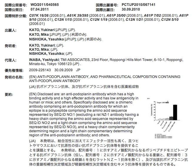
*抗ポドプラニン抗体(NZ-1)の特許が公開になりました。
(WO/2011/040565) 抗ポドプラニン抗体、及び抗ポドプラニン抗体を含む医薬組成物
発明の名称:
(EN) ANTI-PODOPLANIN ANTIBODY, AND PHARMACEUTICAL COMPOSITION CONTAINING ANTI-PODOPLANIN ANTIBODY
(JA) 抗ポドプラニン抗体、及び抗ポドプラニン抗体を含む医薬組成物
要約:
(EN) Disclosed are: an anti-podoplanin antibody which has a high binding activity and a high effecter activity and has low antigenicity in human or mice; and others. Specifically disclosed are: a chimeric antibody comprising an anti-podoplanin antibody for which an epitope is a polypeptide comprising the amino acid sequence represented by SEQ ID NO:1 (excluding a rat NZ-1 antibody having a heavy chain comprising the amino acid sequence represented by SEQ ID NO:2 and a light chain comprising the amino acid sequence represented by SEQ ID NO:3), and a heavy chain complementarity determining region and a light chain complementary determining region of the anti-podoplanin antibody; and others.
(JA)本発明は、結合活性に加えてエフェクター活性も高く、且つヒトやマウスにおいて抗原性の低い抗ポドプラニン抗体等を提供することを課題とする。 本発明は、配列番号:1に示すアミノ酸配列からなるポリペプチドをエピトープとする抗ポドプラニン抗体(但し、配列番号:2に示すアミノ酸配列からなる重鎖と、配列番号:3に示すアミノ酸配列からなる軽鎖とを含むラットNZ-1抗体を除く)、及び当該抗ポドプラニン抗体の重鎖相補性決定領域及び軽鎖相補性決定領域を含むキメラ抗体等を提供するものである。
*NZ-1抗体の特許の公開について
NZ-1抗体の特許はすでにWIPOから公開となっています。(キメラ型抗ヒトpodoplanin抗体(NZ-8))しかし、日本の特許庁が管理する<特許電子図書館>の検索では、この特許が出てこないため、担当の弁理士の先生にこの件を問い合わせました。以下のように教えて頂きました。
「ご質問の件ですが、この特許の出願はPCT出願の国際段階というステージにありますので、日本の特許庁ではなく、WIPOに係属していることになります。公報の公開もWIPOの国際事務局が行っていますので、WIPOのウェブサイトでしか検索できません。 この後、最初の出願日(2009年9月30日)から30ヶ月以内に、権利化を希望する国を選択し、選択した国の特許庁に翻訳文等の書類を提出して、PCT出願を各国の国内段階に移行する手続を行います。この後、出願は各国の特許庁に係属することになり、それぞれの言語で公開されます。
日本の特許庁は、移行後数ヶ月程度で再公表公報と呼ばれる公報を発行します。再公表公報が発行され次第、特許電子図書館で検索できます。再公表公報の発行時期は、あまり一定しておりませんが、特許電子図書館で検索できるようになるのは、2012年の後半ではないかと思います。」
このように、特許のシステムは素人にはとても難しく、毎回勉強になります。研究者も特許の知識がないと、今後の自由な研究ができないような時代が来ると言われています。私もポドプラニンの特許をモデルケースとしては、今後も勉強を続けて行きます。
・Akiko Kunita, Takeshi G. Kashima, Atsushi Ohazama, Agamemnon E. Grigoriadis, Masashi Fukayama
Podoplanin Is Regulated by AP-1 and Promotes Platelet Aggregation and Cell Migration in Osteosarcoma.
The American Journal of Pathology, Volume 179, Issue 2 , Pages 1041-1049 , August 2011
*我々の論文5報(Kato, JBC 2003, Kunita, AJP. 2007, Suzuki, FEBS 2008, Ogasawara, Hybridoma 2008, Kato, Cancer Sci 2008)が引用されている。NZ-1抗体も使用。
Abstract
Podoplanin is a type-I transmembrane sialomucin-like protein, which is expressed in a wide range of cell types and is involved in platelet aggregation and tumor metastasis. Here, we investigated the function, regulation, and expression of podoplanin in osteosarcoma. Podoplanin expression was observed in three osteosarcoma cell lines (MG-63, HOS, and U-2 OS) with platelet aggregation?inducing ability, which was blocked by podoplanin small-interfering RNA or a neutralizing antibody. Overexpression of podoplanin in nonmetastatic Dunn osteosarcoma cells promoted cell migration without attenuating cell proliferation. Both podoplanin and TGF-β1 were up-regulated by c-Fos induction in MC3T3-E1 osteoblastic cells, and were highly expressed in c-Fos transgenic mouse osteosarcomas and c-Fos?transformed osteosarcoma cell lines. Immunohistochemistry of human osteosarcoma tissue microarrays (n = 133) showed staining of tumor cells embedded in an excess of irregular neoplastic bone matrix in 100% of tumors undergoing so-called “normalization/maturation.” Podoplanin was also expressed in osteosarcoma subtypes, with 65% of osteoblastic, 100% of chondroblastic, and 79% of fibroblastic tumors. CD44 and pERM immunohistochemistry showed coexpression with podoplanin in both mouse and human osteosarcoma. Podoplanin expression was significantly higher in metastatic osteosarcomas (n = 6) than in primary osteosarcomas (n = 10). Our data suggest that podoplanin, which is not expressed in normal osteoblasts but in osteocytes, is aberrantly expressed in transformed osteoblasts and in osteosarcoma, and is under AP-1 transcriptional control. Thus podoplanin is a candidate molecule for therapeutic targeting.
*ヒトTh17細胞の選択的分化、同定および調節
特許関連資料
ヒトTh17細胞の分化および活性の両方の調節のための実施形態が提供される。より具体的には、ヒトTH17細胞分化は、TGF-βおよびIL-21、ならびにそれらのアゴニストおよびアンタゴニストによって調節され得る。TH17細胞の機能は、例えば、BLT1もしくはポドプラニン、ならびにそれらのアゴニストおよびアンタゴニストによって調節され得る。さらに、TH17細胞の同定のための実施形態が提供される。より具体的には、ヒトTH17細胞は、BLT1およびポドプラニンを特異的にアップレギュレートする。
・Honma M, Minami-Hori M, Takahashi H, Iizuka H.
Podoplanin expression in wound and hyperproliferative psoriatic epidermis: Regulation by TGF-β and STAT-3 activating cytokines, IFN-γ, IL-6, and IL-22.
J Dermatol Sci. 2011 Dec 8. [Epub ahead of print]
*我々の論文(Suzki, FEBS lett 2008)が引用されています。 NZ-1抗体も使用されています。
Abstract
Podoplanin (PDPN)/T1α/aggrus/PA2.26 antigen, a transmembranous glycoprotein, is a well-known lymphatic endothelial marker. Recent evidence indicates that PDPN is also expressed in keratinocytes especially of sebaceous glands. OBJECTIVE: To verify expression-pattern and the regulatory mechanism of PDPN in human epidermal keratinocytes. METHODS: PDPN-expression pattern was analyzed in normal and psoriatic epidermis by immunostaining. The regulatory mechanism of PDPN-expression of keratinocytes by cytokines was analyzed using specific inhibitors, siRNA, and adenoviral shRNA of signaling pathways. RESULTS: In normal skin, PDPN was expressed on the basal cell layer of sebaceous glands and on the outer root sheath of hair follicles. While no expression was detected in the normal interfollicular epidermis, PDPN was detected in the basal cell layer of wound and hyperproliferative psoriatic epidermis, where the granular layer is lacking. TGF-β1 and IFN-γ independently upregulated PDPN-expression of keratinocytes via TGF-β receptor-Smad pathway and JAK-STAT pathway, respectively. IL-6 and IL-22 also stimulated PDPN-expression of keratinocytes accompanied by STAT-3 phosphorylation. siRNA of STAT-1, inhibitors of STAT-3 signaling, AG490, STAT-3 inhibitor VI, and si/shRNA of STAT-3 inhibited the PDPN-expression of keratinocytes induced by IFN-γ, IL-6 and IL-22 but not by TGF-β1. CONCLUSION: These results indicate that TGF-β1, IFN-γ, IL-6, and IL-22 induce PDPN-expression of keratinocytes, which might be significantly involved in the wound healing process as well as in the pathomechanism of hyperproliferative psoriatic epidermis.
・Finney BA, Schweighoffer E, Navarro-Nunez L, Benezech C, Barone F, Hughes CE, Langan SA, Lowe KL, Pollitt AY, Mourao-Sa D, Sheardown S, Nash GB, Smithers N, Reis E Sousa C, Tybulewicz VL, Watson SP.
CLEC-2 and Syk in the megakaryocytic/platelet lineage are essential for development.
Blood. 2011 Dec 20. [Epub ahead of print]
*いつものWatsonのグループの論文です。我々の論文(Suzki-Inoue, JBC 2007)が引用されています。
Abstract
The C-type lectin receptor CLEC-2 signals through a pathway that is critically dependent on the tyrosine kinase Syk. We show that homozygous loss of either protein results in defects in brain vascular and lymphatic development, lung inflation and perinatal lethality. Furthermore, we find that conditional deletion of Syk in the haematopoietic lineage, or conditional deletion of CLEC-2 or Syk in the megakaryocyte/platelet lineage, also causes defects in brain vascular and lymphatic development, although the mice are viable. In contrast, conditional deletion of Syk in other haematopoietic lineages had no effect on viability or brain vasculature and lymphatic development. We show that platelets, but not platelet releasate, modulate the migration and intercellular adhesion of lymphatic endothelial cells through a pathway that is dependent on CLEC-2 and Syk. These studies demonstrate that megakaryocyte/platelet expression of CLEC-2 and Syk is required for normal brain vasculature and lymphatic development and that platelet CLEC-2 and Syk directly modulate lymphatic endothelial cell behaviour in vitro.
・Peters A, Pitcher LA, Sullivan JM, Mitsdoerffer M, Acton SE, Franz B, Wucherpfennig K, Turley S, Carroll MC, Sobel RA, Bettelli E, Kuchroo VK.
Th17 Cells Induce Ectopic Lymphoid Follicles in Central Nervous System Tissue Inflammation.
Immunity. 2011 Dec 14. [Epub ahead of print]
*注目していたTH17/podoplaninの論文がとうとう出ました(Immunity; IF:24)。我々の論文(Suzki-Inoue, JBC 2007)が引用されています。
Abstract
Ectopic lymphoid follicles are hallmarks of chronic autoimmune inflammatory diseases such as multiple sclerosis (MS), rheumatoid arthritis, Sjogren's syndrome, and myasthenia gravis. However, the effector cells and mechanisms that induce their development are unknown. Here we showed that in experimental autoimmune encephalomyelitis (EAE), the animal model of MS, Th17 cells specifically induced ectopic lymphoid follicles in the central nervous system (CNS). Development of ectopic lymphoid follicles was partly dependent on the cytokine interleukin 17 (IL-17) and on the cell surface molecule Podoplanin (Pdp), which was expressed on Th17 cells, but not on other effector T cell subsets. Pdp was also crucial for the development of secondary lymphoid structures: Pdp-deficient mice lacked peripheral lymph nodes and had a defect in forming normal lymphoid follicles and germinal centers in spleen and lymph node remnants. Thus, Th17 cells are uniquely endowed to induce tissue inflammation, characterized by ectopic lymphoid follicles within the target organ.
・Pula B, Jethon A, Piotrowska A, Gomulkiewicz A, Owczarek T, Calik J, Wojnar A, Witkiewicz W, Rys J, Ugorski M, Dziegiel P, Podhorska-Okolow M.
Podoplanin expression by cancer-associated fibroblasts predicts poor outcome in invasive ductal breast carcinoma.
Histopathology. 2011 Dec;59(6):1249-1260. doi: 10.1111/j.1365-2559.2011.04060.x.
*我々の論文3報(Kato, JBC 2003, Kato, Tumor Biol. 2005, Mishima, ANP 2006)が引用されている。
Abstract
Pula B, Jethon A, Piotrowska A, Gomulkiewicz A, Owczarek T, Calik J, Wojnar A, Witkiewicz W, Rys J, Ugorski M, Dziegiel P & Podhorska-Okolow M (2011) Histopathology 59, 1249-1260 Podoplanin expression by cancer-associated fibroblasts predicts poor outcome in invasive ductal breast carcinoma Aims:? It has recently been shown that podoplanin, a mucin-type glycoprotein, is expressed by cancer cells and cancer-associated fibroblasts (CAFs), and promotes cancer cell migration and invasiveness. The biological role of podoplanin expression in tumour stroma of invasive ductal carcinoma of the breast (IDC) has not been determined. Methods and results:? Podoplanin expression was analysed in 117 cases of IDC and 27 cases of fibrocystic change, as well as in breast cancer cell lines, with the use of immunohistochemistry and real-time polymerase chain reaction. In 82.1% of analysed tumours, podoplanin was found only in CAFs. Only two of 117 IDC cases (1.7%) were characterized by expression of this glycoprotein in cancer cells. None of the fibrocystic changes or stroma surrounding normal ducts showed podoplanin expression. Podoplanin-positive CAFs correlated with tumour size (P?=?0.0125), grade of malignancy (P?=?0.0058), lymph node metastasis (P?=?0.0149), lymphovascular invasion (LVI) (P?=?0.0486) and Ki67 expression in cancer cells (P?=?0.0128). High-level podoplanin expression (>50% of positive stroma) in the tumour stroma was significantly associated with a negative oestrogen status (P?=?0.0201). Univariate, but not multivariate, analysis showed that podoplanin expression by CAFs was associated with poor patient outcome (P?=?0.0202). Conclusions:? Our results suggest that podoplanin expression by CAFs could be an unfavourable prognostic marker for IDC.